Recurrence in #12 (7) after Treatment of 2 Big Cysts
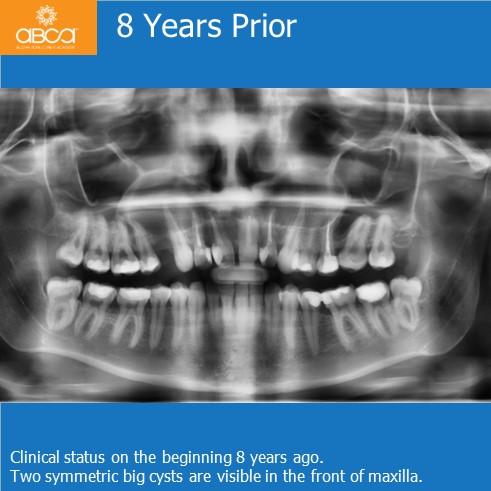
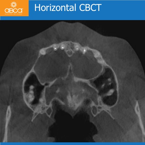
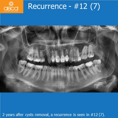
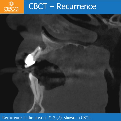
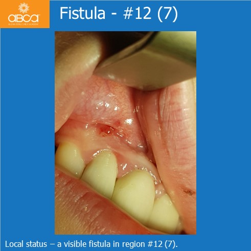
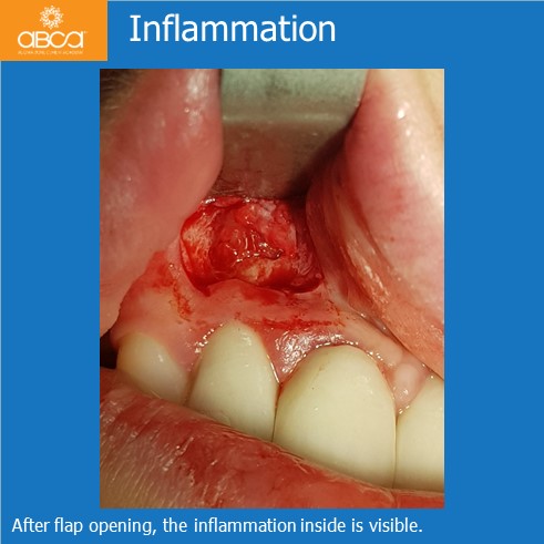
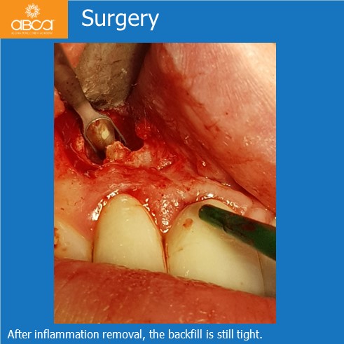
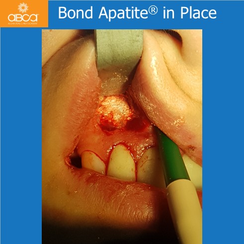
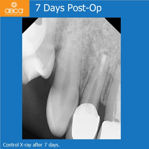
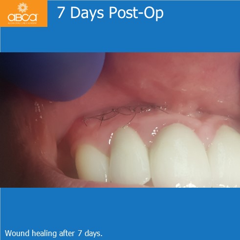
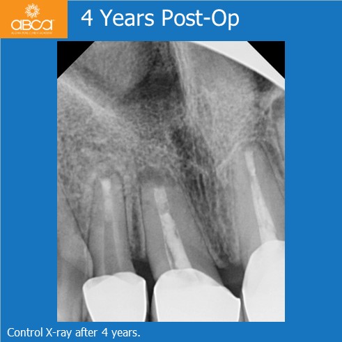
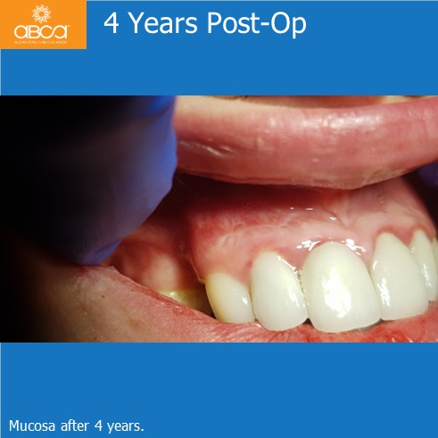
The patient is a healthy, 22-year-old female. Two huge cysts appeared in a panoramic seven years ago, both located in the front of maxillary bone. There were no clinical symptoms. Treatment involved the surgical removal of both cysts, and root resections in teeth #12 (7), #11 (8), #21 (9), #22 (10). Root canals were filled with MTA, but the bone defects were left empty and only L-PRF was inserted. Histopathology showed radicular cysts. 2 years later the patient came back with mild swelling and pain in the right side. In the area of #12 (7) a fistula was found, and secondary inflammation appears in X-rays. Another surgery was performed to remove the fistula. After removing the inflammation, it was noted that the backfill of root canal #12 (7) was still tight. This time the bone defect was filled with Bond ApatiteŽ (1 cc). Good healing and no clinical symptoms were observed throughout a 4-year follow-up period.
Moreover, and most important, is the second histopathological examination which showed inflammation chronic fibrosis. This means that there was no bacteria leakage and recurrence of the radicular cyst, but rather reparative connective tissue inside the bone. This is what happens when defects are left empty, no matter their size!
