Expansive Lesion of the Right Maxilla Extraction and Immediate Reconstruction with Augma Bond ApatiteŽ
The patient is a 60-year-old, healthy, female who came to the clinic for a consultation regarding palatal swelling. She reported a slow growing pattern of the lesion for 4-5 months, which was not painful and had no bleeding or pus discharge during this time.
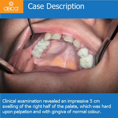
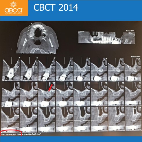
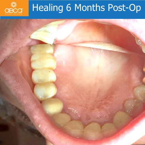
Clinical examination revealed an impressive 5 cm swelling of the right half of the palate, which was hard upon palpation and with gingiva of normal colour. The patient had been to the clinic before for an implant insertion and right sinus lift in 2014. The primary thought was that our challenge is a nonodontogenic lesion of the palate.
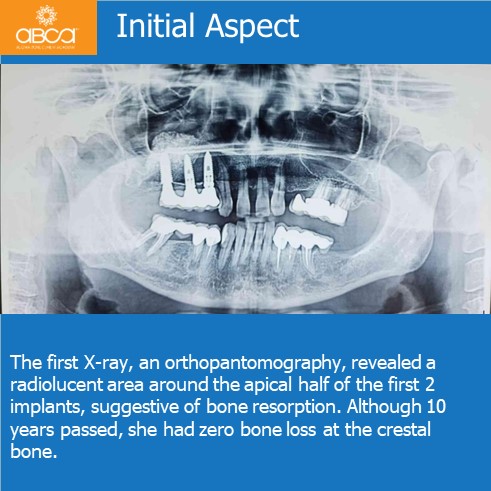
The first X-ray, an orthopantomography, revealed a radiolucent area around the apical half of the first 2 implants, suggestive of bone resorption. Although 10 years passed, she had zero bone loss at the crestal bone.
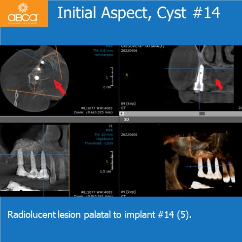
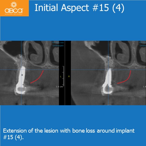
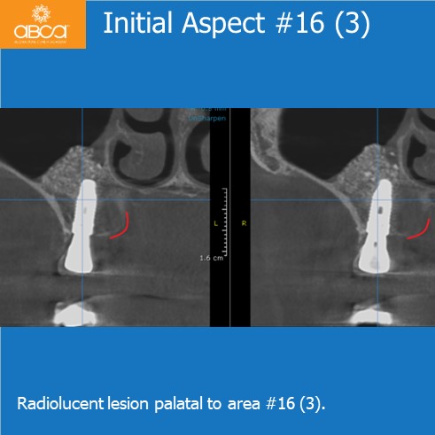
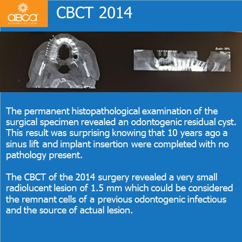
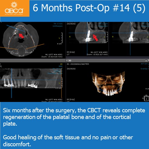
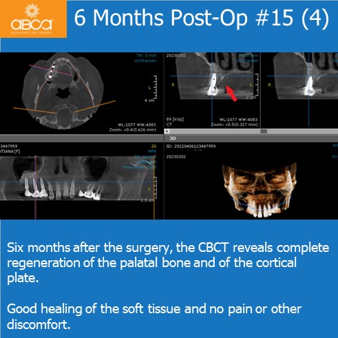
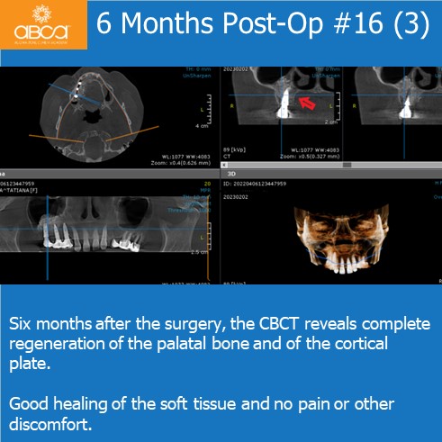
The CBCT showed us the extension of the lesion, which completed eroded the cortical plate over the middle implant and expanded the cortical plate of the other two. There was no contact with the maxillary sinus and no sinusitis.
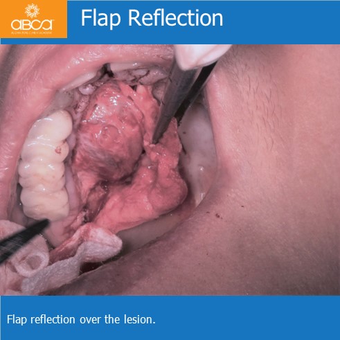
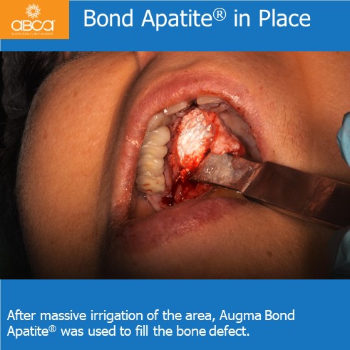
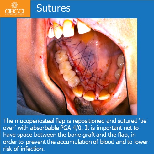
An operation was planned to resect the lesion completely and reconstruct the palatal bone with Bond ApatiteŽ. The patient was taken to surgery, where local anaesthesia was administered. An incision was made on the palatal mucosa, parallel but 5 mm away from the gingival margin, with great consideration for the mucosal adaptation of the crowns. A mucoperiosteal flap was reflected very gently through subperiosteal dissection to expose the lesion and the right maxilla. Care was taken not to tear the membrane of the lesion and the gingiva, to prevent dehiscence and compromise the healing.
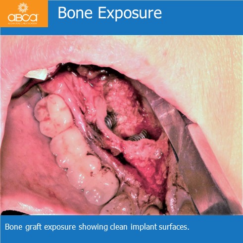
The lesion was removed very easy from the bone and surface of the implants, leaving the area clean. The bone graft from the previous sinus lift was visible, and was clean, hard, and had no granulated tissue and no sinus communications.
At that moment it was clear that the lesion was benign.
